Key Takeaways
- Coelom refers to a true body cavity lined entirely by mesodermal tissue, providing space for organ development and movement.
- Haemocoel is an open circulatory system cavity found in certain invertebrates, mainly arthropods and mollusks, lacking complete mesodermal lining.
- The structural differences between coelom and haemocoel influence how organs are supported and how fluids circulate within the body.
- While coeloms enable compartmentalized organ systems, haemocoels facilitate a less specialized, more fluid-based body plan in invertebrates.
- Understanding these cavities is essential for comprehending the evolutionary adaptations of different animal groups.
What is Coelom?
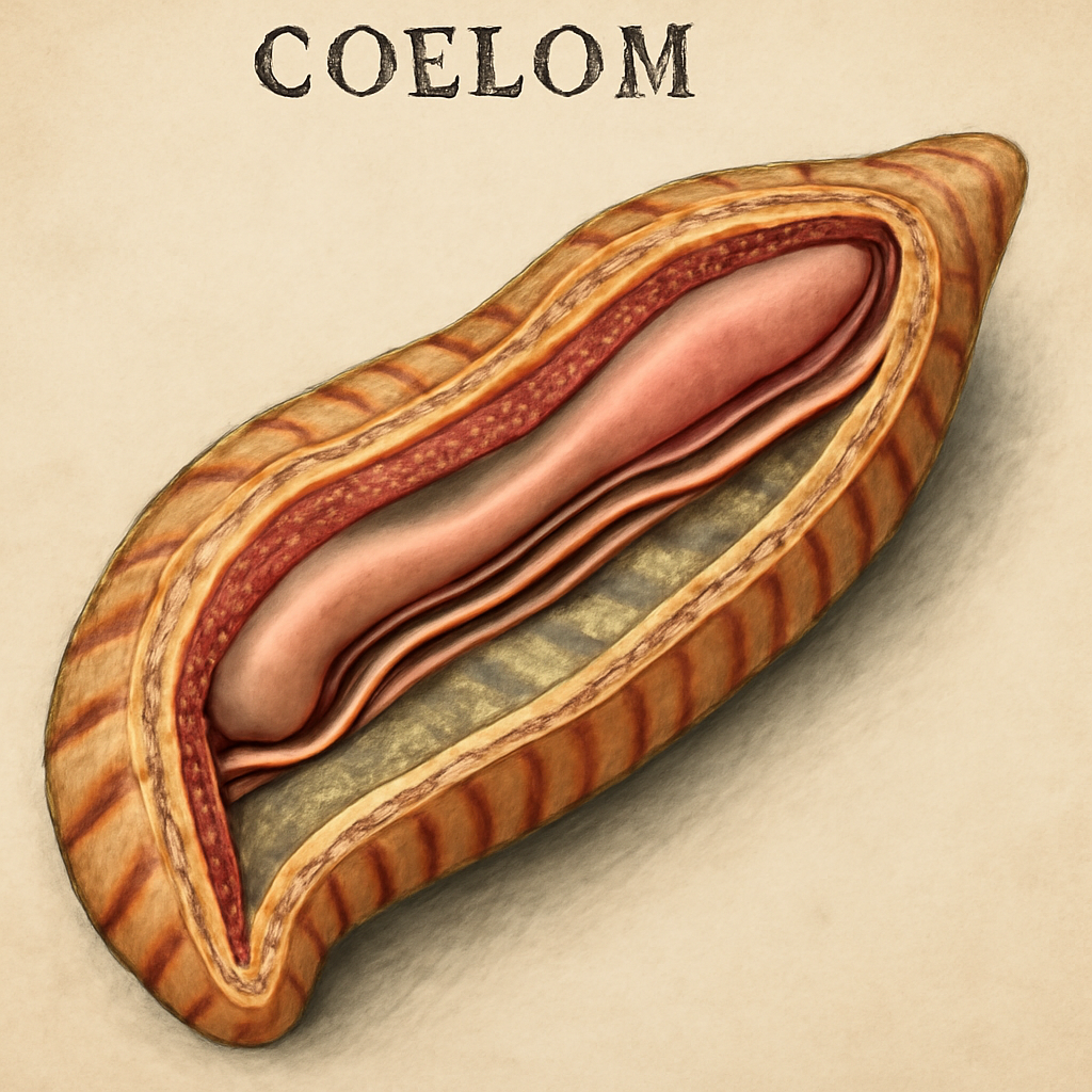
The coelom is a fluid-filled body cavity that is entirely lined with mesodermal tissue, forming a true coelom. It appears during embryonic development in many animals, particularly in vertebrates and some invertebrates, providing a space for organ growth and movement,
Formation of the Coelom During Embryogenesis
The formation of the coelom occurs through a process called schizocoely or enterocoely, depending on the animal’s lineage. In schizocoely, mesodermal masses split to create the cavity, whereas in enterocoely, pouches of mesoderm pinch off from the primitive gut. Although incomplete. This cavity begins as a simple slit that gradually expands to accommodate developing organs.
In vertebrates, the coelom forms during early embryonic stages, creating distinct compartments that later differentiate into thoracic and abdominal cavities. The process involves complex signaling pathways, which ensure proper separation and organization of the mesodermal tissues.
In some invertebrates like annelids, the coelom develops as a segmented cavity, supporting their metameric body plan. The formation process is crucial for the body plan’s structural integrity and functional compartmentalization.
This developmental process allows animals to regulate internal pressure, facilitate movement, and provide a protective cushion for vital organs. The coelom’s presence is a defining feature in many phyla, influencing their morphology and physiology.
Structural Composition and Role in Organ Support
Structurally, the coelom is a spacious, fluid-filled cavity lined with mesodermal epithelium, which supports the internal organs. This lining, called the peritoneum in many cases, secretes lubricating fluids that allow organs to move smoothly during bodily functions.
The coelom acts as a hydraulic skeleton in some animals, enabling movement and maintaining shape by hydraulic pressure. It also serves as a buffer against mechanical shocks, protecting sensitive tissues and organs from external forces.
In vertebrates, the coelom houses vital organs such as the heart, lungs, intestines, and liver, each supported by mesenteries—double layers of peritoneum that contain blood vessels, nerves, and lymphatics.
Additionally, the coelom allows for the independent growth of organs, meaning that organs can develop without constraining each other, facilitating complex organ systems. This separation is crucial in maintaining physiological functions and efficient nutrient distribution across organs.
Evolutionary Significance and Variations
The presence of a coelom marks an important evolutionary step in the complexity of animal body plans. It distinguishes coelomates from acoelomates and pseudocoelomates, reflecting different levels of structural organization.
In evolutionary terms, coeloms have been associated with increased mobility, better organ protection, and the development of a more efficient circulatory system. These advantages contributed to the diversification of many animal groups, especially vertebrates.
Variations among coelomates include different types of coeloms, such as schizocoelous and enterocoelous coeloms, each with distinct developmental origins. These differences influence the body segmentation and organ arrangement in various species.
The coelom’s evolutionary adaptability are evident in how different animals have optimized its structure for their ecological niches, from the segmented worms to complex mammals. Its presence reflects a high level of organismal complexity and specialization.
Functional Implications in Reproductive and Excretory Systems
The coelom provides space for the development and functioning of reproductive organs, allowing for the growth of gonads and related structures without impeding other bodily functions. In many animals, the coelom also facilitates the transport of gametes and nutrients to reproductive tissues.
In excretory systems, the coelom supports the placement and operation of nephridia or kidneys that regulate waste removal. The fluid-filled cavity ensures that waste products are efficiently transported and expelled, maintaining internal homeostasis,
In vertebrates, the coelom’s compartments allow for the separation of circulatory and reproductive systems, reducing cross-interference and optimizing function.
Moreover, the coelom’s fluid dynamics assist in buffering internal pressures during reproductive cycles, which can involve significant changes in organ size and position, especially in gravid females.
This cavity’s role in supporting reproductive and excretory system development underscores its importance for organismal health, growth, and reproduction strategies.
What is Haemocoel?
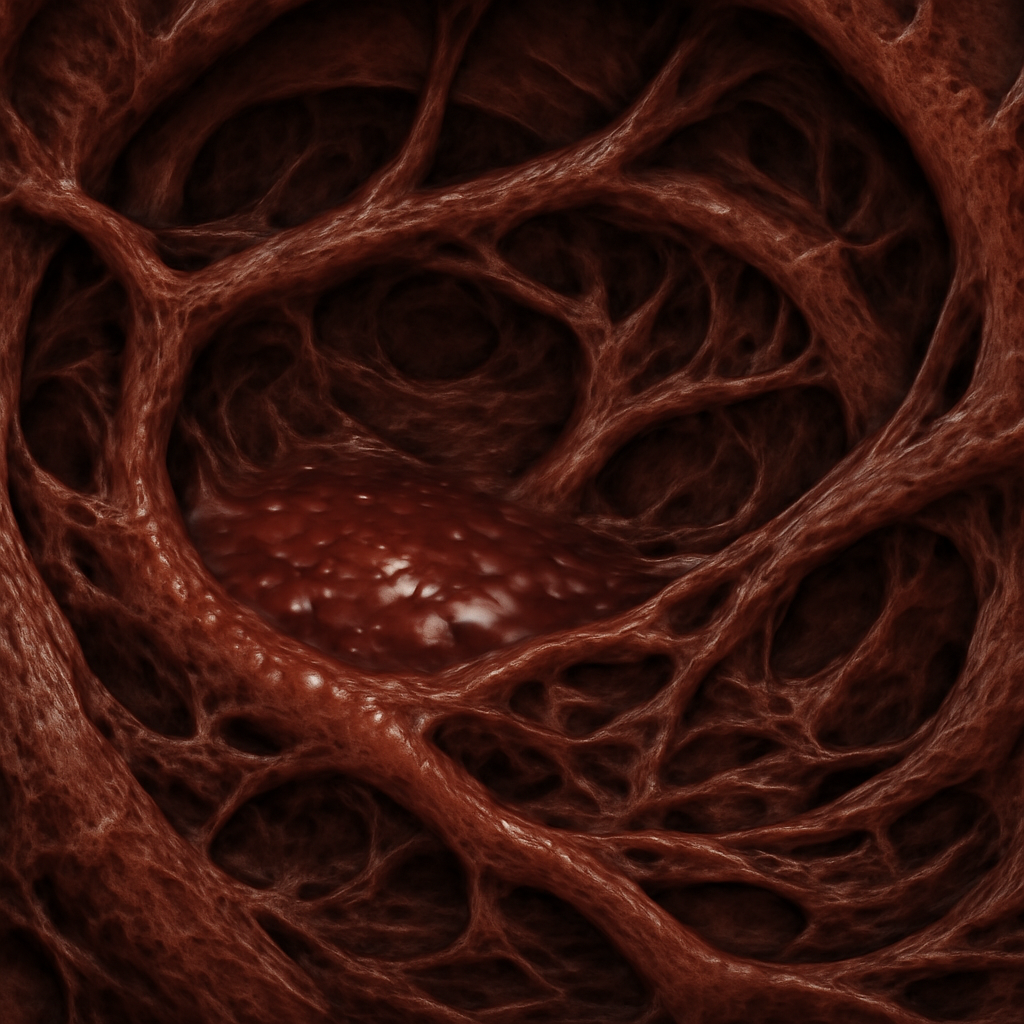
The haemocoel is a body cavity filled with hemolymph, functioning as part of an open circulatory system found primarily in invertebrates like insects, crustaceans, and mollusks. Unlike coeloms, it lacks a complete mesodermal lining and is not a true cavity in developmental terms.
Formation and Evolutionary Context of the Haemocoel
The haemocoel develops as an open space where hemolymph circulates freely, bathing organs directly without the confinement of vessels. It originated as an adaptation in invertebrates to support their metabolic needs and body structures.
During evolution, the haemocoel replaced closed circulatory systems in certain groups, providing a less energy-intensive way to distribute nutrients and remove waste products. This adaptation allowed for larger body sizes and increased mobility in some invertebrates.
The formation of the haemocoel involves the expansion of the coelomic cavity or the development of a new space that is not fully mesoderm-lined. It often coexists with remnants of embryonic coeloms, but it is functionally distinct.
This cavity’s development reflects a different evolutionary pathway, favoring simplicity and efficiency over compartmentalization seen in coelomates. It highlights an alternative strategy for internal support and fluid distribution in animals.
Structural Composition and Fluid Dynamics
The haemocoel is filled with hemolymph, a fluid that combines aspects of blood and interstitial fluid, circulating through open sinuses and cavities. The absence of vessel walls means that hemolymph flows freely, driven by muscular movements and pressure gradients.
Hemolymph transports nutrients, hormones, and waste products directly to and from organs, providing nutrients and removing metabolic byproducts. Its circulation is less directed compared to closed systems, relying on body movements to facilitate flow.
The structural components include sinuses and spaces formed by tissues, which are lined by a thin layer of epithelial cells. This setup allows hemolymph to bathe organs directly, facilitating exchange processes.
In insects, the haemocoel is particularly well-developed, supporting their high activity levels and metabolic demands. Its design simplifies the circulatory system but limits the ability to regulate blood flow precisely.
Functional Roles in Invertebrate Physiology
In invertebrates, the haemocoel functions as a hydrostatic skeleton, providing structural support and enabling movement. It also plays a crucial role in immune responses, as hemocytes circulate within the haemolymph, fighting infections.
Because the haemocoel allows direct organ exposure to circulating fluids, it facilitates rapid distribution of nutrients and quick response to environmental changes. This system supports the animal’s ability to survive in varied habitats.
The open circulatory system, with the haemocoel as its core, is energy-efficient, reducing the need for complex vascular networks. This efficiency is advantageous for small and medium-sized invertebrates with high activity levels.
Despite its limitations regarding precise control of blood flow, the haemocoel enables invertebrates to maintain effective internal environments despite external fluctuations, contributing to their evolutionary success.
Implications for Organ Development and Movement
The haemocoel allows organs to develop in a less constrained environment, giving flexibility in spatial arrangement. This setup supports the development of large, complex organs in some invertebrate species.
Movement is aided by the hydrostatic pressure generated by hemolymph, which acts as a supportive fluid, allowing animals like insects to jump, crawl, or fly efficiently. The haemocoel’s fluid dynamics directly influence locomotion and posture.
In terms of organ function, the direct exposure to hemolymph means that tissues are more accessible for immune cells and nutrients, making physiological responses faster and more integrated.
This cavity’s design influences the overall body plan, favoring versatility and adaptability, especially in organisms that undergo rapid or energetic movements.
Understanding the haemocoel’s structure elucidates how invertebrate species have evolved to maximize efficiency with minimal structural complexity.
Comparison Table
Below is a detailed comparison based on structural, developmental, functional, and evolutionary aspects:
| Parameter of Comparison | Coelom | Haemocoel |
|---|---|---|
| Type of Body Cavity | True cavity lined with mesoderm | Open cavity filled with hemolymph |
| Developmental Origin | Formed via schizocoely or enterocoely | Develops as a spacious sinuses and spaces, often from coelomic remnants |
| Line of Lining | Mesodermal epithelial lining | Lacks complete mesodermal lining, exposed tissues |
| Circulatory System Type | Closed circulatory system (in many animals) | Open circulatory system with hemolymph circulation |
| Support for Organs | Supports and suspends organs via mesenteries | Supports organs through hydrostatic pressure and tissue contact |
| Fluid Content | Contains coelomic fluid, separate from blood or hemolymph | Filled with hemolymph, functions as blood equivalent |
| Evolutionary Significance | Associated with advanced organ system development | Linked to simpler, energy-efficient body plans in invertebrates |
| Body Flexibility | Provides structural rigidity and compartmentalization | Allows high flexibility and movement with less rigidity |
| Examples of Organisms | Vertebrates, some invertebrates like annelids | Insects, mollusks, arthropods |
| Role in Reproductive Systems | Supports gonads and reproductive organs with space for development | Less involved, as reproductive organs are often embedded directly in tissues |
| Protection Against Mechanical Shock | Provides cushioning for internal organs | Offers limited protection, relies more on external exoskeletons |
Key Differences
Here are some specific points that distinguish Coelom and Haemocoel:
- Structural lining — coeloms are fully lined with mesodermal tissue, whereas haemocoels are not, making the latter less compartmentalized.
- Developmental origin — coeloms originate from embryonic processes like schizocoely or enterocoely, while haemocoels develop as spaces within tissues, often from coelomic remnants.
- Circulatory function — coeloms support closed circulatory systems, whereas haemocoels are associated with open circulatory systems where hemolymph flows freely.
- Support for organs — in coeloms, organs are suspended and supported by mesenteries; in haemocoels, organs are bathed directly in hemolymph, relying on hydrostatic pressure.
- Organ development — coeloms facilitate complex organ development with separate compartments; haemocoels allow more flexible, less compartmentalized organ arrangements.
- Fluid content — coeloms contain coelomic fluid, while haemocoels are filled with hemolymph, which functions as blood in invertebrates.
- Evolutionary role — coeloms are linked to advanced body plans, whereas haemocoels are characteristic of simpler, energy-efficient invertebrate systems.
FAQs
How does the presence of a coelom influence the animal’s mobility?
The coelom provides a hydrostatic skeleton that enhances structural support and allows for more precise and controlled movements, especially in segmented animals like worms and vertebrates, by acting as a flexible yet sturdy internal cavity.
Can an organism have both coelom and haemocoel at the same time?
Yes, some invertebrates possess remnants of coelomic cavities alongside haemocoels, with the former supporting certain organ systems and the latter facilitating circulation, reflecting an evolutionary transition or functional specialization.
What are the implications of having a haemocoel for immune responses?
The haemocoel allows hemolymph to circulate freely, carrying immune cells that respond quickly to infections or injuries, providing an efficient means of defense in invertebrates lacking a closed circulatory system.
How does the structure of the coelom impact organ differentiation?
The internal space of the coelom supports the development of specialized, well-organized organs by providing enough room and protective lining, which is crucial for complex organ systems seen in vertebrates and some invertebrates.